
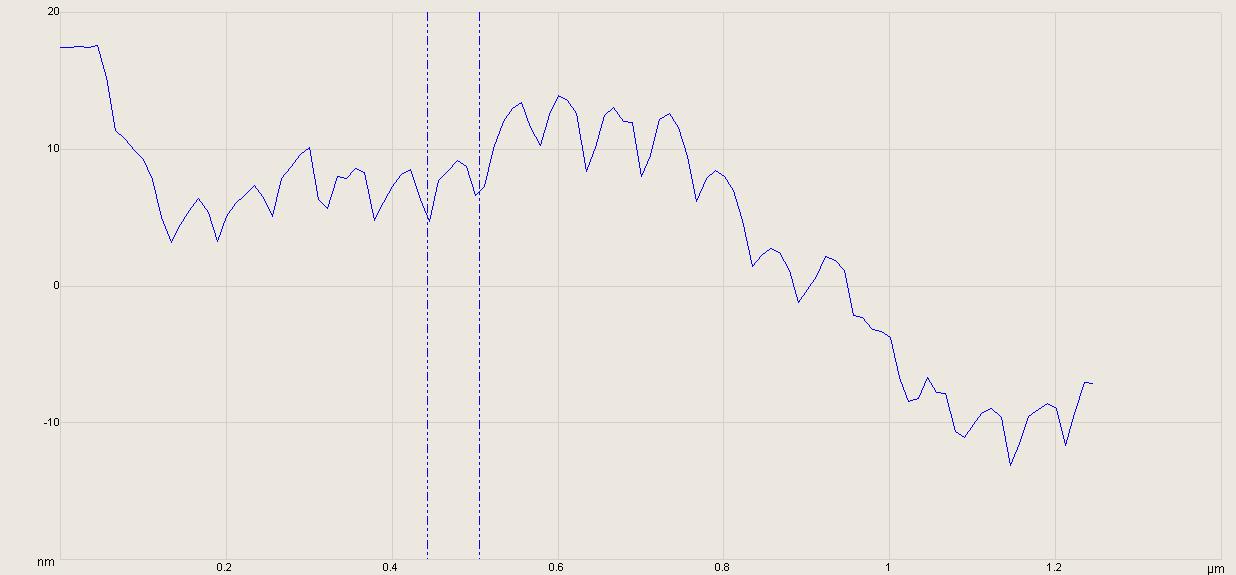
 The D-bands arising from the staggering of tropocollagen structural units is clearly visualized on individual collagen fibrils. By drawing the sectioning tool across individual fibers, an effective digital cross section of the fiber along that line is produced. In this case the spacing between two specific bands is found to be 54 nm. While the D-band period is a function of such environmental parameters as hydration, this is far less than the expected 67 nm seen in TEM of stained fibrils.
Application of the sectioning tool can be problematic as measurements are then biased according to the operator's selection of targets and placement of the dimensioning cursors. It is human nature of select features that are the least ambiguous and simplest to dimension by interacting with the image. As an example in this case I chose to section a long fiber in a cluster of long fibers. Removing operator bias would require a sampling methodology which covered more of the image field, including less "attractive" looking fibers, and statistically combining these measurements.
No comments:
Post a Comment