Showing posts with label flattening. Show all posts

Wednesday, August 1, 2018

Image Sectioning: Collagen D-bands (Part 1)

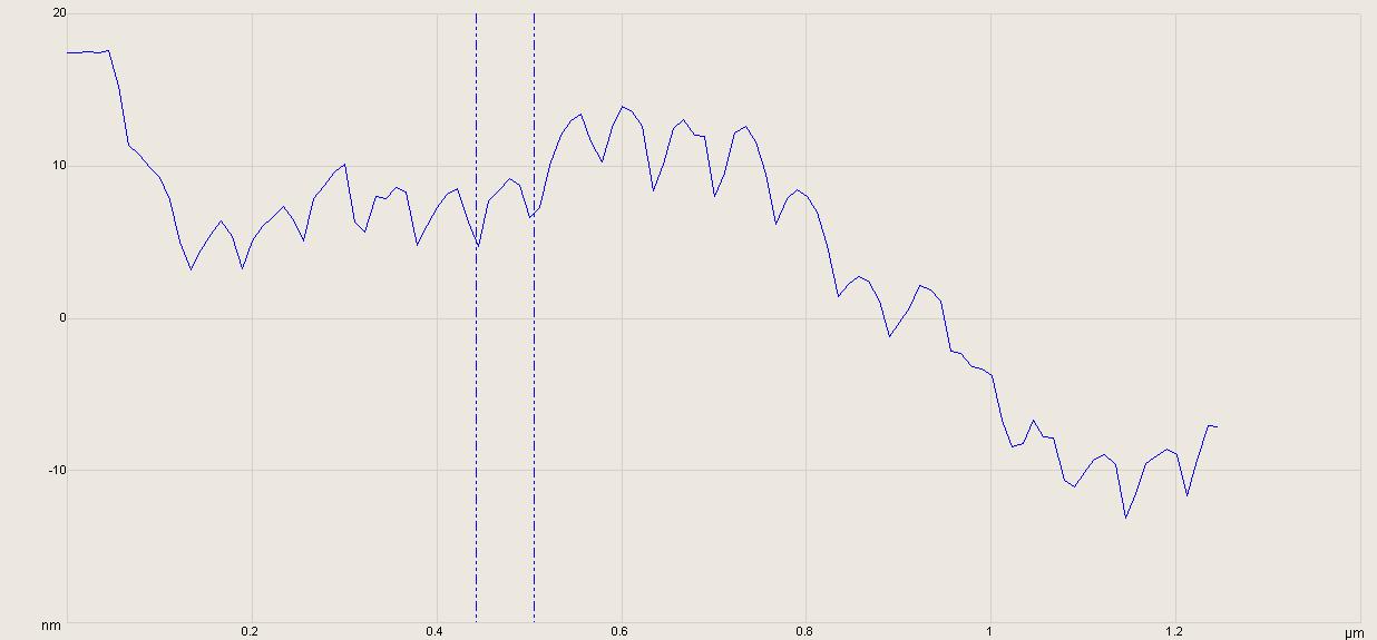
Images can be sectioned using the sectioning tool. In this case an image of human collagen was taken with PeakForce tapping. The image was flattened to remove large scale spatial relationships not related to the structure of individual collagen fibrils.

The D-bands arising from the staggering of tropocollagen structural units is clearly visualized on individual collagen fibrils. By drawing the sectioning tool across individual fibers, an effective digital cross section of the fiber along that line is produced. In this case the spacing between two specific bands is found to be 54 nm.  While the D-band period is a function of such environmental parameters as hydration, this is far less than the expected 67 nm seen in TEM of stained fibrils.

Application of the sectioning tool can be problematic as measurements are then biased according to the operator's selection of targets and placement of the dimensioning cursors. It is human nature of select features that are the least ambiguous and simplest to dimension by interacting with the image. As an example in this case I chose to section a long fiber in a cluster of long fibers. Removing operator bias would require a sampling methodology which covered more of the image field, including less "attractive" looking fibers, and statistically combining these measurements.

Thursday, June 30, 2011

Flattening & Flattening Artifacts

AFM images generally require some post-processing. AFM images are generated by scaling the Z-piezo voltages to yield a height, and that voltage generally has an offset required to extend or retract the Z-piezo so that the probe can interact with the sample. There is often also a tilt to the sample due to mounting or the cleaving of the substrate which is irrelevant to the surface morphology but nevertheless reflected in the AFM image. The offset and tilt prevent a meaningful interpretation of the AFM image. Such an image is shown at top.


One method of post-processing is flattening. In this case a polynomial of order n is fit and subtracted from every trace and retrace. It is called "flattening" because this method of processing tends to flatten or unroll an AFM image. It is also a potential source of artifacts.

The second image shows a series of photo-lithographed Au pads for directed self-assembly. Flattening was applied in image post-processing. Note the dark regions extending in the fast scan direction on either side of the pads. In AFM any features that are consistently and uniformly aligned in the fast direction are suspect. According to the flattened image the substrate near the pads dips down-- but only on the right and left sides of the pads. This is physically implausible and suspect. Any sections, height, bearing or Ra measurements from this image would be erroneous.

To remedy these artifacts stop bands are placed on the pads allowing only the substrate pixels to be used in the flattening. In this final image stop bands are placed over the pads in the middle portion of the image showing a flattened image without baseline distortions.

In general, flattening works best post processing images that don't have high aspect ratio features: e.g. grains on a film surface. In these applications one is interested in the small scale structure, not the large scale morphology. Flattening long wavelength features away is in fact often desirable. In cases like these lithographed pads flattening can still be applied but beware of artifacts and use stop bands accordingly.